YBioTech Research Trend · Cell Imaging

Cytation C10 도입 전
확인해야 할 5가지

Confocal과 Widefield 자동 이미징, Live Cell 분석, 선택 구성에 따른 multimode microplate detection까지. Cytation C10은 단순히 “현미경 한 대”를 고르는 문제가 아니라 연구 workflow 전체를 어떻게 설계할 것인지 함께 보는 장비입니다.
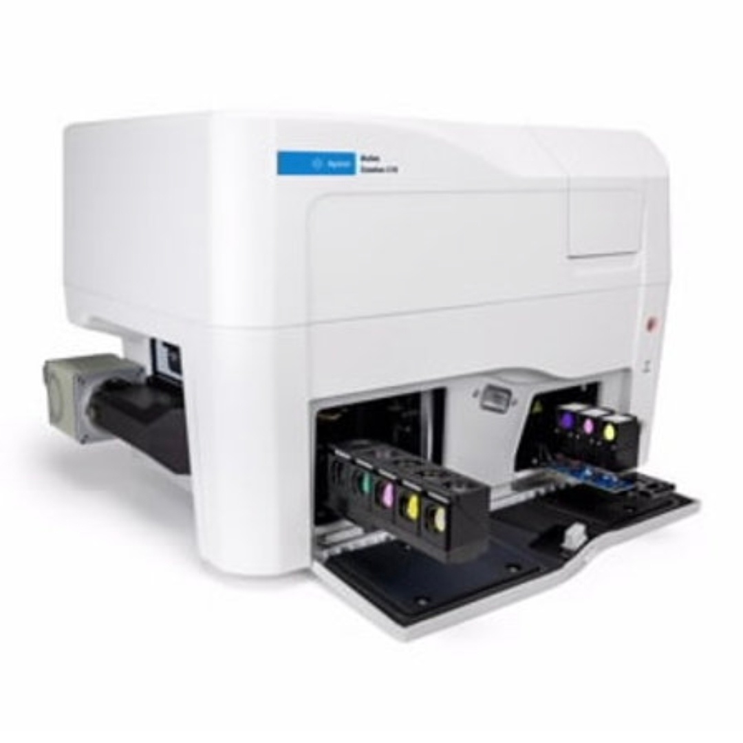
Spinning Disk Confocal Widefield Imaging Water Immersion Live Cell Gen5 Analysis Multimode Detection
Agilent BioTek Cytation C10
Confocal두꺼운 시료의 optical sectioning
Widefield빠른 이미지 획득과 screening
Water Immersion고배율 수침 이미징 지원
Live Cell환경 제어 기반 kinetic imaging
Multimode선택 구성에 따른 plate detection
BEFORE SELECTION

장비보다 먼저
실험을 확인하세요.

Cytation C10은 automated confocal microscopy와 widefield microscopy를 하나의 플랫폼에서 운용하고, 필요에 따라 conventional multimode microplate reading까지 결합할 수 있는 imaging reader입니다.

하지만 2D monolayer cell을 빠르게 확인하는 실험과 organoid·spheroid 같은 두꺼운 3D sample의 내부 구조를 분석하는 실험은 필요한 광학 조건이 다릅니다. 장시간 Live Cell assay라면 온도와 CO₂/O₂ 환경, 시간 간격, plate 처리량까지 함께 고려해야 합니다.

핵심 포인트
“C10에 어떤 기능이 있는가?”보다 먼저 “우리 연구에서 무엇을 보고, 무엇을 정량화해야 하는가?”를 정리하면 필요한 구성과 데모 조건을 훨씬 명확하게 잡을 수 있습니다.
From Sample to Data

C10 하나로 연결되는 연구 흐름

단순 이미지 획득에서 끝나는 것이 아니라, 3D sample의 구조 확인부터 시간에 따른 변화와 정량 분석까지 하나의 workflow로 설계할 수 있습니다.

3D SAMPLE

Organoid · Spheroid

두꺼운 시료에서 관심 초점면과 내부 형광 구조를 더 선명하게 확인해야 하는 연구.
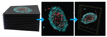
OPTICAL SECTIONING

Confocal + Z-stack

여러 Z-plane을 획득하고 3D biology의 depth 정보를 활용하는 imaging workflow.
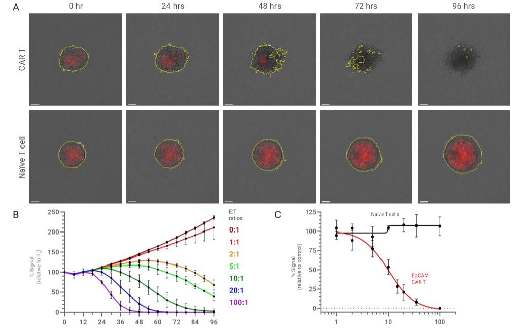
QUANTIFICATION

Image → Gen5 → Data

세포 수, 면적, 형광 강도, object 기반 지표 등 반복 가능한 정량 분석으로 연결.

Cytation C10 Technology

C10의 핵심 기술을 먼저 이해하세요

Cytation C10은 confocal, widefield, high-quality optical components, environmental control 및 Gen5 기반 분석을 하나의 시스템 안에서 연결합니다.

Widefield Fluorescence · Brightfield · Phase Contrast

모든 이미지를 confocal로 촬영할 필요는 없습니다. 빠른 plate survey, cell confluence 및 일반적인 fluorescence imaging에는 widefield optics가 효율적일 수 있습니다.
  • Widefield fluorescence imaging
  • Brightfield imaging
  • Phase contrast imaging

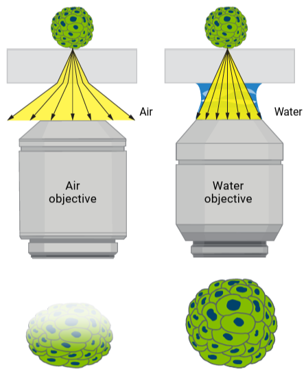
Water Immersion Objectives

Cytation C10은 자동화된 water immersion imaging을 지원하여 높은 배율에서 수침 대물렌즈를 이용한 imaging workflow를 구성할 수 있습니다.
  • 40x / 60x water immersion 지원 구성
  • 고해상도 cellular detail 관찰
  • 고처리량 automated imaging과 결합

Scientific CMOS Camera · Laser Illumination

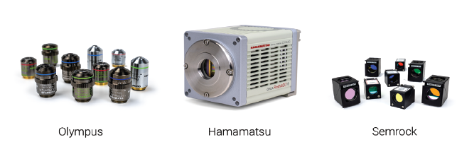
Agilent는 Cytation C10에 Hamamatsu scientific CMOS camera, Olympus objectives 및 laser-based illumination을 사용하는 고품질 optical platform으로 설명하고 있습니다.

  • sCMOS 기반 image acquisition
  • 고품질 microscope objectives
  • laser-based illumination
Official Spec Snapshot

도입 검토 시 자주 보는 주요 사양

실제 발주 구성은 옵션에 따라 달라질 수 있으므로 최종 사양은 견적 단계에서 확인하세요.

AIR OBJECTIVES 1.25x–60x Air objective 구성 범위
PHASE CONTRAST 4x–40x Phase contrast magnification range
CONFOCAL 20x–60x Confocal magnification range
WATER IMMERSION 40x / 60x Water immersion objective options
DETECTION Abs · FI · LUM 선택 구성에 따른 multimode detection
CO₂ CONTROL 0–20% Optional Gas Controller 구성 시
O₂ CONTROL 1–19% Optional Gas Controller 구성 시
SOFTWARE Agilent BioTek Gen5 Imager / reader control & analysis
5 Things to Check

도입 전 확인해야 할 5가지

사양표를 먼저 보는 것보다 실제 실험 workflow를 기준으로 아래 5가지를 정리해 보세요.

CHECK01
SAMPLE TYPE

어떤 시료를 관찰할 것인가?

2D monolayer인지, spheroid·organoid 같은 3D model인지, tissue나 biofilm처럼 두꺼운 sample인지 먼저 정리해야 합니다. 시료의 두께와 관찰 깊이는 widefield와 confocal 선택에 직접적인 영향을 줍니다.

POINT현재 시료뿐 아니라 향후 2~3년의 주요 sample까지 함께 정리하세요.
CHECK02
IMAGING MODE

Widefield와 Confocal을 어디에서 나눠 쓸 것인가?

빠른 전체 plate 확인과 일반적인 cellular imaging에는 widefield가 효율적일 수 있고, 두꺼운 3D sample의 특정 깊이·내부 구조·Z-stack 분석에서는 confocal의 필요성이 커집니다.

POINT“모든 이미지를 confocal로 찍는다”가 아니라 workflow 단계별 역할을 정하세요.
CHECK03
LIVE CELL

시간에 따른 세포 변화를 봐야 하는가?

proliferation, migration, cytotoxicity, cell-cell interaction 및 drug response처럼 시간 정보가 중요한 assay라면 endpoint image보다 kinetic imaging이 중요해집니다. 장시간 측정에서는 온도와 CO₂/O₂ 환경, imaging interval과 총 실험 시간을 함께 봐야 합니다.

POINTEndpoint / 몇 시간 / 며칠 단위 assay인지 먼저 구분하세요.
CHECK04
IMAGE ANALYSIS

이미지를 보는 것에서 끝나는가, 숫자로 바꿔야 하는가?

Gen5를 이용하면 image capture, processing 및 analysis workflow를 연결할 수 있습니다. Cell count, object area, morphology, fluorescence intensity 등 최종 endpoint를 먼저 정의하면 acquisition 조건도 더 체계적으로 설계할 수 있습니다.

POINT“예쁜 이미지”보다 최종적으로 필요한 정량 지표를 먼저 적어보세요.
CHECK05
FUTURE WORKFLOW

Imaging 외에 plate reader와 automation까지 확장할 것인가?

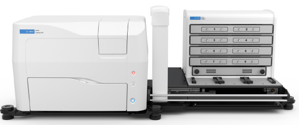
Cytation C10은 선택 구성에 따라 absorbance, fluorescence intensity, luminescence microplate detection을 함께 사용할 수 있습니다. 또한 BioSpa 8 Automated Incubator와 연계하면 multiplate live-cell workflow로 확장할 수 있습니다.

POINT현재 실험만 보지 말고 screening, automation, plate detection 확장 가능성까지 검토하세요.
Research Applications

C10이 특히 잘 맞는 연구 workflow

Agilent의 Cytation C10 application 자료에서 반복적으로 다뤄지는 연구 영역을 도입 관점에서 정리했습니다.

3D

Organoid · Spheroid

두꺼운 3D sample의 confocal sectioning, Z-stack 및 구조 분석.

LC

Live Cell Imaging

세포의 증식, 이동, 형태 변화와 약물 반응을 시간에 따라 반복 관찰.

IO

Immuno-Oncology

3D tumor model, immune-cell interaction 및 cell killing kinetics 연구.

BF

Biofilm Imaging

두꺼운 bacterial biofilm의 confocal imaging과 구조 기반 평가.

NV

Neurobiology

신경세포 및 복잡한 생물학적 구조의 automated imaging workflow.

HS

High Content Screening

다수 well의 자동 이미지 획득과 반복 가능한 정량 분석.

DR

Drug Response

약물 처리 전후 phenotype, viability 및 fluorescence signal 변화 분석.

RT

Retinal / Tissue Imaging

조직과 같이 복잡하고 두꺼운 sample의 detail과 depth 정보 관찰.

MM

Imaging + Multimode

형태학적 image와 plate-based quantitative signal을 하나의 workflow에서 활용.

Selection Workflow

C10 도입 검토는 이렇게 시작하세요

시료와 endpoint를 먼저 정리하면 필요한 광학 구성과 옵션이 자연스럽게 좁혀집니다.

STEP 01

Sample

2D / 3D / tissue / biofilm 등 실제 분석 시료와 두께를 정리합니다.

STEP 02

Imaging

Widefield / Confocal / Z-stack / magnification과 채널 조건을 정합니다.

STEP 03

Analysis

Cell count, morphology, intensity 등 최종 quantitative endpoint를 정의합니다.

STEP 04

Expansion

Live Cell 환경, multimode detection, BioSpa 8 automation까지 향후 확장을 검토합니다.

Gen5 Software

이미지 획득에서 분석까지 Gen5로 연결

Gen5는 BioTek imaging 및 microplate reader workflow에서 image capture, processing, analysis 및 data collection을 통합하는 소프트웨어 플랫폼입니다.

CONTROL · PROCESS · ANALYZE

이미지를 찍는 것보다
분석 workflow가 중요합니다.

실험마다 동일한 acquisition 조건과 analysis setting을 반복 적용하고, image 기반 endpoint를 데이터로 정리해야 high-content workflow의 장점을 살릴 수 있습니다.
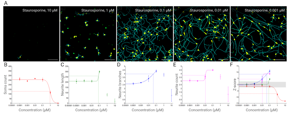
Image Acquisition Image Processing Object Analysis Fluorescence Quantification Protocol Automation Data Export
Live Cell & Automation

한 plate를 넘어 multiplate live-cell workflow로

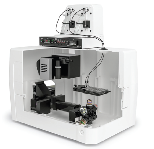
Cytation C10의 onboard environmental control은 live-cell assay를 지원하며, BioSpa 8 Automated Incubator와 연계하면 여러 plate의 장시간 자동 workflow로 확장할 수 있습니다.
Onboard Environmental Control

장시간 live cell imaging에서는 세포 상태를 안정적으로 유지하는 것이 중요합니다. 실험 조건에 따라 temperature, CO₂/O₂ control 필요성을 함께 검토해야 합니다.
FAQ

C10 도입 검토 시 자주 묻는 질문

오가노이드 연구라면 반드시 Confocal이 필요한가요?
연구 목적에 따라 다릅니다. 전체 크기와 morphology를 빠르게 보는 목적과, 내부 특정 구조·깊이별 signal을 분리해 보는 목적은 필요한 imaging 방식이 다릅니다. 실제 sample 두께와 관찰 endpoint를 기준으로 데모 조건을 잡는 것이 좋습니다.
Widefield와 Confocal을 한 protocol에서 나눠 사용할 수 있나요?
가능합니다. 빠른 survey에는 widefield를 사용하고 관심 well이나 두꺼운 sample을 confocal로 정밀 분석하는 식으로 workflow를 설계할 수 있습니다.
Water immersion objective는 어떤 연구에서 고려하나요?
높은 배율에서 cellular detail을 더 정밀하게 관찰해야 하는 경우 고려할 수 있습니다. 실제 sample type, plate, required resolution과 throughput을 함께 봐야 합니다.
Live Cell Imaging을 할 때 무엇을 먼저 정해야 하나요?
총 assay 시간, imaging interval, required magnification, fluorescence exposure, temperature와 CO₂/O₂ 필요성, plate 수를 먼저 정리하는 것이 좋습니다.
이미지 분석은 Gen5에서 할 수 있나요?
Gen5는 image acquisition, processing 및 analysis workflow를 통합합니다. 필요한 endpoint에 따라 object-based analysis와 quantitative setting을 사전에 설계할 수 있습니다.
Plate Reader 기능도 동시에 사용할 수 있나요?
Cytation C10은 구성에 따라 UV-Vis absorbance, fluorescence intensity, luminescence detection을 포함한 multimode microplate reading 기능을 사용할 수 있습니다.
도입 전에 실제 시료로 데모하는 것이 좋을까요?
가능하다면 실제 연구에 사용할 sample, plate, fluorescence label, magnification, imaging mode와 최종 analysis endpoint를 기준으로 테스트하는 것이 장비 적합성을 판단하는 데 도움이 됩니다.
Agilent Official Resources

더 깊게 확인하고 싶다면

아래 자료는 Cytation C10의 기술, 사양과 application을 확인할 때 참고하기 좋은 Agilent 공식 자료입니다.

Cytation C10 Product Page Confocal + Widefield + multimode platform의 제품 개요와 주요 기능. Agilent 공식 페이지 →
Cytation C10 Technical Details Magnification, detection mode, environmental control 등 상세 사양. Agilent Technical Details →
Confocal Technology Spinning disk confocal 기술과 Cytation C10 confocal imaging 원리. Confocal Technology →
Gen5 Software for Imaging & Microscopy Image capture, processing, analysis를 통합하는 Gen5 software 정보. Gen5 공식 페이지 →
Water Immersion Imaging Cytation C10의 automated water immersion imaging 관련 공식 application 자료. Application Note →
Agilent Image Gallery Cytation C10을 포함한 Agilent BioTek imaging examples 확인. Image Gallery →

내 연구에는 어떤 Cytation C10 구성이 필요할까요?

사용 예정 시료, plate, fluorescence label, 필요한 magnification과 imaging mode, Live Cell 여부 및 최종 분석 endpoint를 알려주시면 도입 검토에 필요한 구성을 함께 정리할 수 있습니다.